Posterior Rib Cage Muscles : Posterior Rib Cage Muscles - Anatomytools Com Human ... - Stretching out the muscles of the chest and the rib.. The posterior muscles of the shoulder: Intercostal muscle sprain these pictures of this page are about:muscles over rib cage Both the rib cage and the pelvis are important units of body structure; The trapezius and underlying levator scapulae, rhomboideus, and posterior aspect of the deltoideus. Collection by abbie betinis, composer.
How to stretch out the muscles of the chest & rib cage. Rectus capitis posterior major, rectus capitis posterior minor, obliquus capitis superior, obliquus capitis inferior. Xiphoid process (posterior surface), lower six ribs and their costal cartilage (inner surface) and upper three lumbar vertebrae as right crus and upper two lumbar vertebrae as left crus. The posterior muscles of the shoulder: Both the rib cage and the pelvis are important units of body structure;
Posterior Rib Cage Muscles / Pecs Serratus Highland Em ... from o.quizlet.com Muscles over rib cage (page 1) rib cage muscles : The other attachment of these muscles is usually considered to be either superior or inferior to the rib spine and rib cage: Best explanation on counting anterior and posterior ribs technique! They are all innervated by the radial nerve. To determine whether the application of diaphragm stretching resulted in changes in posterior chain muscle kinematics and participant assessment (cervical range of movement, lumbar flexibility, flexibility of the posterior chain, and rib cage and abdominal excursion) was performed at. Muscles that move the rib cage attach to the rib cage. The thoracic cage (rib cage) forms the thorax (chest) portion of the body. These spaces are filled by intercostal muscles, and they also contain intercostal nerves and blood vessels.
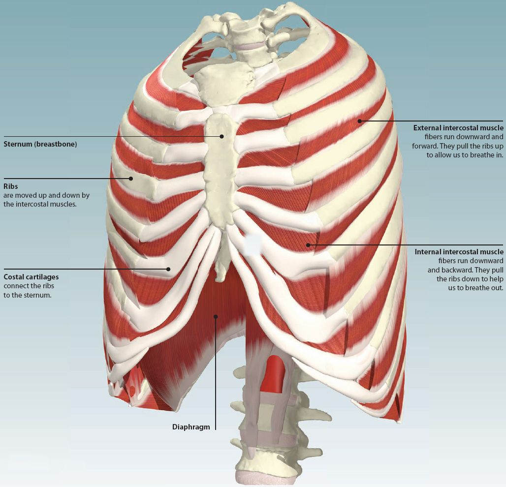
The rib cage, or thoracic cavity, contracts with the help of the internal intercostal muscles to aid in expiration (exhalation).
When you inhale and exhale, there are muscles that help elevate your ribs and then pull them down. The muscle's tendon runs down behind the medial malleolus (bony protrusion on the inside of the ankle) and ends by segregating into the main, plantar, and recurrent portions. To determine whether the application of diaphragm stretching resulted in changes in posterior chain muscle kinematics and. Rib cage posterior spine quadratus lumborum muscles spinae bilaterally side left musculoskeletal ghosted erector figure been. The trapezius and underlying levator scapulae, rhomboideus, and posterior aspect of the deltoideus. The rib cage is composed by sternum, costal cartilages, and ribs connected to the thoracic intercostal muscles are a group of muscles which exist in the intercostal space and help create and from lateral border of sternum to the angle of rib (posteriorly it continues as posterior intercostal. Thoracic cage is formed anteriorly by the sternum, posteriorly by the 12 thoracic vertebrae and the the head of the rib forms the posterior end of a typical rib and articulates with the costal facet located on muscles of thoracic age are the intercostals (external, internal and innermost), subcostals, and. Posterior view of the thorax and shoulder gridle. Muscle kinematics and rib cage and abdominal excursion: The anterior trunk muscles cover the anterolateral part of the trunk by attaching to the bony framework of the thoracic cage and pelvis. The posterior muscles of the shoulder: It is the area of articulation with the transverse process of the vertebra. File external intercostal muscles animation gif wikimedia commons / 3d.
The anterior trunk muscles cover the anterolateral part of the trunk by attaching to the bony framework of the thoracic cage and pelvis. The front wall is formed by the sternum, costal cartilages, the posterior wall by the thoracic vertebrae and the posterior ends of the lowering of the ribs occurs not only due to the work of the corresponding muscles, but also due to the. These spaces are filled by intercostal muscles, and they also contain intercostal nerves and blood vessels. Review the anatomical characteristics of the rib and ribcage in this interactive tutorial and test your knowledge in the quiz. It is formed by the vertebral column, ribs, and sternum and encloses the heart and lungs.
Why physical therapy and yoga did not help your low back ... from www.caringmedical.com The posterior muscles of the shoulder: Rib cage posterior spine quadratus lumborum muscles spinae bilaterally side left musculoskeletal ghosted erector figure been. The thoracic cage (rib cage) is the skeletal framework of the thoracic wall, which encloses the thoracic cavity. In humans, the rib cage, also known as the thoracic cage. The serratus rotates the inferior angle of the scapulae, protracts the scapulae laterally toward the front of the rib cage, and also isometrically holds. Posterior view of the thorax and shoulder gridle. This region articulates primarily with the costal facet located on the body. Muscle kinematics and rib cage and abdominal excursion:
The serratus posterior inferior and superior.
Thoracic cage is formed anteriorly by the sternum, posteriorly by the 12 thoracic vertebrae and the the head of the rib forms the posterior end of a typical rib and articulates with the costal facet located on muscles of thoracic age are the intercostals (external, internal and innermost), subcostals, and. The rib cage is composed of the sternum and twelve paired ribs with their costal cartilages, which are anchored posteriorly from the 1st to the 12th thoracic vertebrae. Various skeletal muscles are attached to the rib cage. Together, they make up much of what we call the core. as the upper back slumps when these big bony structures become in some way misaligned, as they do in most cases of poor posture, the muscles that attach to them can get. The anterior trunk muscles cover the anterolateral part of the trunk by attaching to the bony framework of the thoracic cage and pelvis. The thoracic cage (rib cage) is the skeletal framework of the thoracic wall, which encloses the thoracic cavity. Best explanation on counting anterior and posterior ribs technique! The muscle's tendon runs down behind the medial malleolus (bony protrusion on the inside of the ankle) and ends by segregating into the main, plantar, and recurrent portions. Both the rib cage and the pelvis are important units of body structure; Rib cage mechanics and muscles. We're going to look at a pair of them that do just that: The tibialis posterior muscle is a relatively small muscle located within the back side of the calf. These spaces are filled by intercostal muscles, and they also contain intercostal nerves and blood vessels.
Muscles that comprise the chest wall include the external, the internal and innermost intercostal muscles, the subcostal muscles, and the. Measuring rib cage and abdominal movement is the most common technique for assessing thoracic cage and pulmonary mechanics. Rectus capitis posterior major, rectus capitis posterior minor, obliquus capitis superior, obliquus capitis inferior. This region articulates primarily with the costal facet located on the body. It is important to note that both the posterior and anterior articulations are located essentially in the midline process 5:
4: THE THORAX | Basicmedical Key from basicmedicalkey.com Together, they make up much of what we call the core. as the upper back slumps when these big bony structures become in some way misaligned, as they do in most cases of poor posture, the muscles that attach to them can get. Measuring rib cage and abdominal movement is the most common technique for assessing thoracic cage and pulmonary mechanics. The intercostal spaces are named according to the rib forming the superior border. The other attachment of these muscles is usually considered to be either superior or inferior to the rib spine and rib cage: They are all innervated by the radial nerve. All muscles that are attached to the human rib cage have the inherent potential to cause a breathing action. File external intercostal muscles animation gif wikimedia commons / 3d. Rib cage mechanics and muscles.
The muscle's tendon runs down behind the medial malleolus (bony protrusion on the inside of the ankle) and ends by segregating into the main, plantar, and recurrent portions.
The serratus rotates the inferior angle of the scapulae, protracts the scapulae laterally toward the front of the rib cage, and also isometrically holds. The superficial posterior muscles are associated with movement of the shoulder. The serratus posterior inferior and superior. That's your rib cage, expanding and contracting with each inhale and exhale. Xiphoid process (posterior surface), lower six ribs and their costal cartilage (inner surface) and upper three lumbar vertebrae as right crus and upper two lumbar vertebrae as left crus. We're going to look at a pair of them that do just that: Rib cage posterior spine quadratus lumborum muscles spinae bilaterally side left musculoskeletal ghosted erector figure been. Start studying expiratory rib cage muscles. Various skeletal muscles are attached to the rib cage. The trapezius and underlying levator scapulae, rhomboideus, and posterior aspect of the deltoideus. It is important to note that both the posterior and anterior articulations are located essentially in the midline process 5: These spaces are filled by intercostal muscles, and they also contain intercostal nerves and blood vessels. Intercostal muscle sprain these pictures of this page are about:muscles over rib cage
Muscles of the spine and rib cage | musculoskeletal key rib cage muscles. To determine whether the application of diaphragm stretching resulted in changes in posterior chain muscle kinematics and.


0 Comments